The Technology Stack
Advanced Cancer Technologies
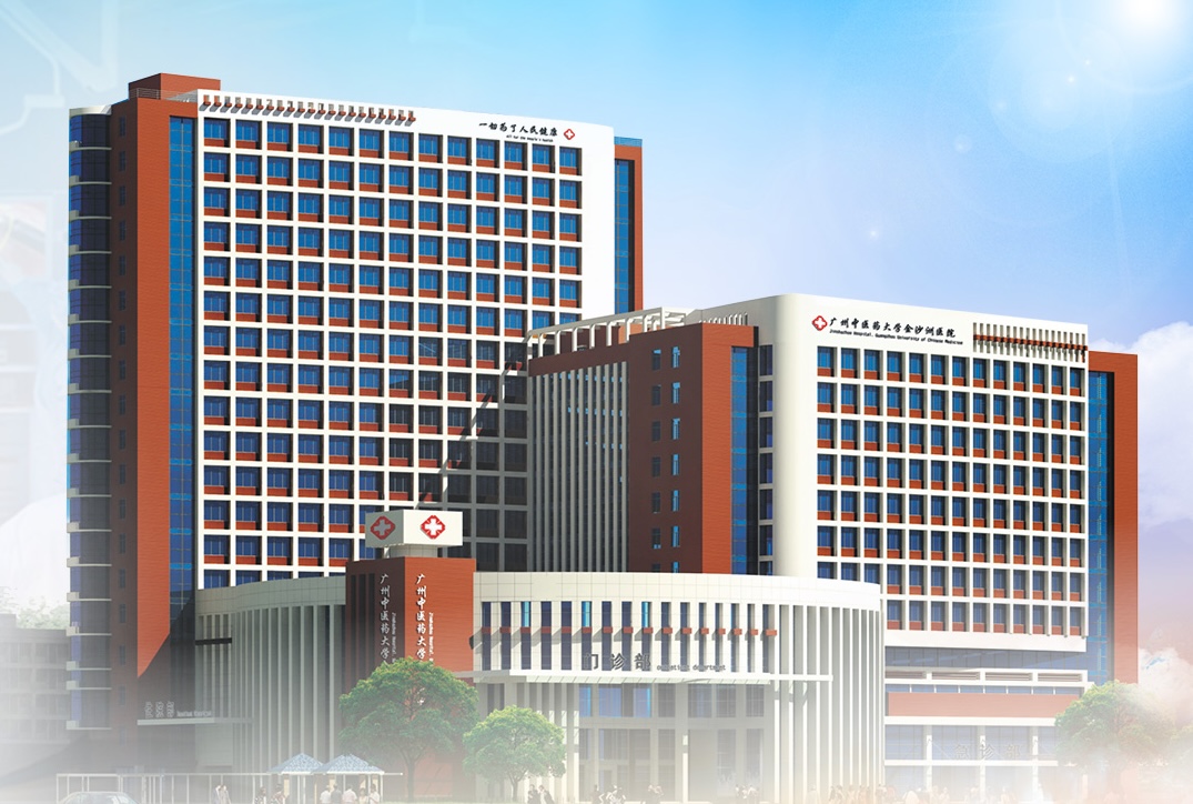
Jinshazhou Hospital of GZUCM provides access to advanced minimally invasive oncology tools — many of which are either unavailable in the Philippines or have waiting lists of months to years.
An AI-driven radiosurgery system with dynamic tracking that delivers high-intensity radiation with sub-millimetre precision, hitting tumours from hundreds of angles while bending beams around healthy anatomy.
The Accuray M6 CyberKnife uses a robotic arm equipped with dynamic tracking capabilities to track tumour movement in real-time as a patient breathes, bending 1,200 radiation beams around anatomy with sub-millimetre accuracy.
A 58-year-old man with a 2.5 cm inoperable lung tumour received three CyberKnife sessions over one week — no incision, no hospital admission. At three months, imaging showed complete tumour response. He flew home without a scar.
The Mevion S250i uses the 'Bragg Peak' effect — charged particles deposit their full energy precisely at tumour depth, then stop, with almost zero radiation beyond the tumour.
Proton therapy exploits the Bragg Peak — unlike X-rays that scatter dose throughout the body, protons deposit their maximum energy precisely at the tumour depth and then stop completely. This significantly reduces radiation to surrounding healthy tissue, lowering the risk of severe complications, IQ decline, and secondary malignancies.
A 9-year-old girl with a brainstem glioma received proton therapy instead of conventional X-ray radiation, protecting her developing brain tissue. Within weeks of finishing treatment, she was back in school.
Radioactive Iodine-125 seeds implanted directly into or around the tumour, delivering continuous low-dose, short-range radiation from within — for months — without systemic exposure.
Radiation Particle Implantation uses tiny radioactive iodine-125 seeds placed under CT guidance directly into the tumour. The seeds silently emit continuous low-dose radiation from the inside, over months, without entering the bloodstream or affecting surrounding tissue.
A 55-year-old man with recurrent lung cancer and limited pulmonary reserve (making re-operation impossible) had a 3 cm tumour in a location where external beam radiation would be imprecise. After iodine-125 seed implantation, the tumour shrank by 80% with no systemic side effects. The seeds remain in place permanently, becoming inert after decay.
Integrates a linear accelerator with a helical CT scanner to deliver 360-degree focal layer irradiation — ideal for complex, multiple, or irregularly shaped tumours.
The TomoTherapy H System combines a linear accelerator with a helical CT scanner on a rotating gantry, allowing it to deliver radiation in a 360-degree spiral pattern around the patient. This approach is particularly effective for tumours with irregular shapes or those that wrap around critical structures, where conventional radiation angles would cause excessive collateral damage.
A 16-year-old female with a grade IV malignant brain tumour (intracranial germ cell tumour) received 25 radiotherapy sessions using TOMO's 360° helical irradiation. Follow-up 10 months later revealed significant shrinkage of her brain lesions.
An image-guided radiotherapy platform that adjusts radiation plans daily as a tumour changes size or shape, with HyperArc technology for treating multiple brain metastases in a single session.
Varian TrueBeam is an IGRT (Image-Guided Radiotherapy) system that captures high-resolution imaging before each session, allowing clinicians to adapt the radiation plan as the tumour shifts or shrinks. Its HyperArc 'Super Arc Knife' technology can treat multiple brain metastases in a single, highly efficient session.
An elderly male patient with lung cancer and multiple brain metastases was confined to a wheelchair with severe intracranial hypertension. After 5 targeted TrueBeam sessions, his tumours regressed, his neurological function was restored, and he was walking independently again.
A high-speed image-guided radiotherapy system delivering simultaneous integrated boost doses — completing treatments up to four times faster than conventional systems.
Varian Halcyon is a ring-gantry IGRT system designed for speed and precision, rotating significantly faster than conventional C-arm linacs to reduce treatment time while maintaining sub-millimetre accuracy. It uses simultaneous integrated boost (SIB) dosing to treat the primary tumour and surrounding at-risk tissue in a single session.
A 49-year-old male diagnosed with diffuse large B-cell lymphoma presenting as a brain lesion was treated with Halcyon in March 2021 using SIB over 20 fractions. Less than a month after finishing treatment, MRI scans confirmed significant reduction of his intracranial lesions.
An AI-powered online adaptive radiotherapy platform that re-contours the tumour and rewrites the treatment plan from scratch before every single session — in real time.
Ethos captures high-definition CT imaging before each treatment session and uses artificial intelligence to instantly re-contour the tumour target and recalculate the full treatment plan on the spot, accounting for daily changes in organ position, shape, and tumour size. This is critical when tumours shift based on organ filling (e.g., bladder, rectum) or when the tumour is actively shrinking during treatment.
A 67-year-old male with bladder cancer had a tumour that changed position daily based on bladder fullness. Using Ethos, the AI re-contoured his target and modified his plan before every session, ensuring radiation hit the tumour precisely despite the organ's fluctuating size.
Irreversible Electroporation uses ultra-short, high-voltage electrical pulses to create permanent holes in tumour cell membranes — without heat, safe next to blood vessels, bile ducts, and nerves.
NanoKnife (Irreversible Electroporation) is a non-thermal technology that utilizes ultra-short, high-voltage electric pulses to create permanent holes in cancer cell membranes, triggering cell death at the molecular level without generating heat. This makes it safe to use right next to critical structures like blood vessels, bile ducts, and nerves that would be damaged by heat-based methods.
A woman with stage 3 pancreatic cancer had a tumour wrapped around her superior mesenteric artery — one of the vessels keeping the intestines alive. Every surgeon said: inoperable. Heat-based ablation would risk the artery. The MDT recommended Nanoknife. Electrical pulses were delivered directly to the tumour. The artery was untouched, the tumour was ablated, and chemotherapy resumed the following week.
Argon-helium cryotherapy uses extreme cold to freeze tumours into an 'ice ball', destroying cancer cells with minimal pain and scarring — through a small probe, often in one session.
Cryoablation (Argon-Helium Knife) uses extreme cold (argon and helium gases) to freeze tumours into an 'ice ball', killing cancer cells completely with minimal pain and scarring. The cryo-knife probe is inserted under ultrasound guidance, and a warming catheter protects adjacent structures like the urethra.
A 70-year-old man with early-stage prostate cancer and multiple comorbidities (heart disease, diabetes, poor renal function) could not safely undergo surgery or radiotherapy. His family had been told to watch and wait. The MDT recommended argon-helium cryotherapy. One session, no general anaesthesia, discharged the same day. PSA levels normalised within three months.
High Intensity Focused Ultrasound converges acoustic energy on the tumour from outside the body, generating heat that destroys it — without incisions, radiation, or anaesthesia.
HIFU (High Intensity Focused Ultrasound) is a completely non-invasive technology that focuses acoustic energy on the tumour from outside the body, heating it to destruction. No cuts, no radiation, no general anaesthesia required.
A 45-year-old woman with a 4 cm liver tumour wasn't opposed to treatment — she was opposed to surgery after watching her father recover from hepatic resection. The MDT offered HIFU, which converges acoustic energy on the tumour from outside the body, destroying it without a single incision. She was discharged the same afternoon and was back at work two days later.
Electromagnetic waves and heat delivered through a thin probe to rapidly destroy tumour tissue — a fast, minimally invasive alternative to surgery for solid tumours.
Microwave Ablation (MWA) and Radiofrequency Ablation (RFA) use electromagnetic energy delivered through a needle-thin probe inserted directly into the tumour under imaging guidance. MWA generates heat through microwave energy for faster ablation of larger tumours; RFA uses alternating electrical current to heat and destroy cancer cells. Both are performed under local anaesthesia or light sedation, require no incision, and can be repeated if needed.
A 58-year-old woman with cirrhosis and three small hepatocellular carcinomas (2–3 cm each) could not withstand major surgical resection. The MDT recommended MWA, performed under ultrasound guidance. All three lesions were ablated in a single 90-minute session with no complications. She was discharged the following day and returned to normal activity within one week.
A catheter threads directly into hepatic arteries, delivering chemotherapy at the source while embolising (cutting off) the blood supply feeding the tumour — without exposing the body to systemic toxicity.
Transarterial Chemoembolisation (TACE) and Hepatic Arterial Infusion Chemotherapy (HAIC) deliver high-dose chemotherapy directly into the arteries feeding liver tumours while simultaneously cutting off their blood supply. A catheter is threaded through the femoral artery directly into the hepatic vessels, concentrating drug delivery at the tumour while embolising the blood vessels that keep the cancer alive.
A 52-year-old man with hepatitis B had three liver tumours (3–6 cm) too numerous and vascular for surgical resection. Systemic chemotherapy would devastate his already struggling liver. After two TACE sessions over eight weeks, CT confirmed necrosis in all three lesions. He was reassessed for curative resection.
Next-Generation Sequencing (NGS) identifies your tumour's genetic mutations — EGFR, ALK, PD-L1, MSI-high — to match you with the most effective targeted drug or immunotherapy regimen.
Targeted and immunotherapy drugs are selected based on your tumour's unique molecular fingerprint, not generic chemotherapy that attacks all fast-growing cells. Biomarker testing identifies actionable mutations (EGFR, ALK, PD-L1, MSI-high) that predict which targeted agents and immunotherapies will work best.
A non-smoking woman in her late 40s was diagnosed with stage 4 lung adenocarcinoma. In the Philippines, her oncologist recommended standard chemotherapy without molecular testing. At GZUCM, NGS revealed an EGFR exon 19 deletion. She was prescribed a third-generation EGFR inhibitor — a drug not yet approved in the Philippines at the time. Within eight weeks, her primary lung tumour shrank by 60% and her bone metastases stabilised. She managed her entire treatment as an outpatient, without hospital admission.
Radioactive microscopic beads delivered directly into the blood vessels supplying liver tumours, destroying cancer cells from within while minimizing damage to healthy tissue.
Advanced nuclear therapy designed specifically for primary liver cancer (HCC) and liver metastases. The microspheres lodge in the tumor's blood supply, delivering high-dose radiation directly at the cancer while sparing the surrounding liver.
A 63-year-old man with advanced HCC and a large (8 cm) primary lesion was deemed inoperable but was a good candidate for bridging therapy before transplantation. After a single Y-90 radioembolisation procedure, imaging at three months showed significant tumour shrinkage (30% reduction), allowing him to proceed to liver transplantation.
A precision nuclear treatment that combines boron-10 compounds with neutron activation to selectively destroy cancer cells at the molecular level.
BNCT represents a frontier in targeted nuclear medicine, offering selective destruction of cancer cells while preserving normal tissue. The hospital is expanding capacity for this emerging technology.
A 52-year-old patient with recurrent malignant glioma had exhausted conventional options (surgery, radiation, chemotherapy) and was facing end-of-life care. After boron-10 accumulation and neutron activation, molecular-level destruction of cancer cells was achieved with minimal damage to surrounding brain tissue, extending survival and quality of life.
Systemic radioisotope treatments targeting specific cancer types — strontium-89 for bone tumours and iodine-131 for thyroid cancer.
These targeted radioisotope therapies deliver therapeutic radiation to cancers with specific uptake characteristics. Strontium-89 concentrates in bone lesions; iodine-131 targets thyroid tissue and thyroid cancer metastases.
A 68-year-old man with prostate cancer and multiple painful bone metastases received strontium-89. Within weeks, his pain score dropped from 8/10 to 2/10, and he regained mobility and independence. A follow-up bone scan six months later showed significantly reduced lesion uptake.
A targeted radioisotope therapy using phosphorus-32 to deliver beta radiation directly to rapidly dividing cancer cells, exploiting their high phosphate uptake.
Phosphorus-32 (32P) is a beta-emitting radioisotope that concentrates in tissues with high metabolic activity — including cancer cells — delivering localised radiation from within. It is used for specific haematological and solid tumour applications where targeted internal radiation is clinically indicated.
A 55-year-old patient with polycythemia vera (a haematological disorder with cancer risk) received phosphorus-32 therapy to reduce excess red blood cell production. The isotope's selective uptake by rapidly dividing bone marrow cells delivered targeted irradiation, normalising blood counts without systemic toxicity.
A frameless radiosurgery system that uses 192 intersecting beams of gamma radiation to destroy brain tumours with extreme precision — without a single surgical incision.
Gamma Knife radiosurgery directs 192 individual beams of cobalt-60 gamma radiation to converge precisely at the tumour target within the brain, delivering a high dose at the intersection while each individual beam passes harmlessly through surrounding tissue. It is a well-established treatment for brain tumours, AVMs, trigeminal neuralgia, and acoustic neuromas, often completing treatment in a single session.
A 71-year-old woman with a 1.5 cm acoustic neuroma causing progressive hearing loss and balance problems was not fit for open neurosurgery. She underwent a single Gamma Knife session. At six-month follow-up, imaging showed no tumour growth, hearing was preserved, and balance deficits resolved.
A high-precision external beam radiotherapy system delivering shaped photon beams to tumours with millimetre accuracy, combining stereotactic targeting with intensity modulation.
Photon Knife therapy uses precisely shaped photon beams in a stereotactic framework to deliver high-dose, highly conformal radiation to tumours throughout the body. The stereotactic approach restricts dose to the target volume while minimising exposure to surrounding healthy structures.
A 44-year-old patient with a small (2 cm) renal cell carcinoma near the renal hilum was not a surgical candidate due to complex vascular anatomy. Photon Knife stereotactic ablative radiotherapy (SABR) delivered four high-dose sessions with millimetre precision. Follow-up imaging at 12 months showed complete local control with preserved renal function.
Internal radiation delivered by placing radioactive sources directly inside or adjacent to the tumour — HDR for high-dose-rate sessions, LDR for continuous low-dose implants.
Brachytherapy uses Varian HDR (High Dose Rate) or LDR (Low Dose Rate) equipment to place radioactive sources directly inside or very close to the tumour. HDR delivers intense, short bursts of radiation through a temporarily placed applicator; LDR uses permanently or temporarily implanted seeds for sustained low-level irradiation. Both approaches deliver maximum dose directly at the tumour while sparing surrounding normal tissue.
A 52-year-old woman with early-stage cervical cancer received four weekly HDR brachytherapy sessions combined with external beam radiotherapy. The radioactive applicator delivered extremely high doses directly to the cervical lesion while protecting the bladder and rectum. She completed treatment as an outpatient with minimal side effects and achieved complete remission.
Highly precise, minimally invasive robotic surgery that allows surgeons to perform complex procedures through tiny incisions with 3D visualization and tremor elimination.
Robotic-assisted surgery offers superior precision, reduced blood loss, and faster recovery compared to traditional open surgery. Ideal for complex cases where traditional techniques would require large incisions.
A 61-year-old man with a 4 cm renal cell carcinoma required partial nephrectomy to preserve kidney function. Da Vinci robotic surgery removed the lesion with millimetre precision through four small (1 cm) port incisions. Hospital stay was one night, pain was minimal, and renal function remained normal at six-month follow-up.
Specialized non-invasive ultrasound technologies that use acoustic energy to destroy tumours without cutting, heating, or radiation.
Advanced acoustic photodynamic systems offer non-invasive destruction of tumours, particularly valuable for patients who cannot undergo surgery or prefer non-surgical options.
A 65-year-old woman with a 3 cm breast cancer near the chest wall declined mastectomy. Using acoustic energy targeted via ultrasound guidance, the lesion was completely destroyed in a single 45-minute outpatient session without general anaesthesia, bleeding, or scarring.
Comprehensive life-support treatments including plasma exchange, bilirubin adsorption, and hemofiltration dialysis for patients with liver dysfunction during cancer treatment.
These advanced supportive care technologies help patients with compromised liver function tolerate aggressive cancer treatments. Critical for patients with hepatitis or cirrhosis undergoing chemotherapy or surgery.
A 58-year-old man with hepatitis B and hepatocellular carcinoma had severely compromised liver function (bilirubin 4.2 mg/dL). Artificial liver support using bilirubin adsorption and plasma exchange reduced bilirubin to 1.8 mg/dL within 48 hours, enabling him to safely proceed with curative-intent chemoembolisation.
Siemens SOMATOM Confidence (dual-energy 4D CT), fourth-generation PET-MR, PET-CT, and 3.0T MRI for precise tumour visualization and staging.
State-of-the-art imaging allows surgeons and oncologists to visualize tumours in three dimensions, identify critical structures, assess metastases, and plan treatment with sub-millimetre accuracy. Four-dimensional imaging captures tumour motion during breathing.
A 58-year-old patient with a lung tumour near the hilum underwent dual-energy 4D CT and PET-MR imaging. The advanced platforms revealed vascular involvement invisible on standard imaging, allowing the MDT to redesign the surgical approach and avoid catastrophic bleeding during resection.
Comprehensive genomic analysis that identifies mutations, biomarkers, and genetic signatures to match each patient with the most effective targeted drugs.
NGS testing identifies actionable mutations (EGFR, ALK, PD-L1, MSI-high, BRCA) that determine which targeted therapies and immunotherapies will work best. Results guide precision treatment selection.
A 42-year-old woman with metastatic colorectal cancer received standard chemotherapy at a local hospital with minimal response. NGS at GZUCM revealed BRAF V600E mutation and high microsatellite instability (MSI-high). She was switched to a BRAF/MEK inhibitor plus immunotherapy combination, achieving rapid tumour shrinkage and improved survival outlook.
Deep genomic analysis that sequences the protein-coding regions of the entire genome to detect rare mutations and predict treatment response.
More comprehensive than standard biomarker panels, WES can uncover unexpected mutations that guide treatment decisions and predict which patients will respond to immunotherapy.
A 48-year-old man with treatment-resistant sarcoma had exhausted conventional options. WES revealed a rare SDHA mutation previously missed by standard panels. This finding opened access to experimental targeted therapies and immunotherapy combinations, leading to a durable response.
⚠️ Note: Not all technologies are available for every cancer type or stage. Eligibility is determined during the MDT case review. The scenarios above are illustrative examples and do not represent guaranteed or typical outcomes. Individual results vary based on cancer type, stage, overall health, and other clinical factors. MediDocPH does not make any promise or guarantee of treatment results.